Galeria
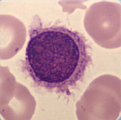
Cell description:
Size: larger than normal lymphocytes
Nucleus: round, oval, dumbbell-shaped or bilobed with little chromatin condensation and sometimes indistinct nucleolus
Cytoplasm: abundant weakly basophilic with irregular “hairy” margins
<p>Cell description: </p> <p>Size: larger than normal lymphocytes </p> <p>Nucleus: round, oval, dumbbell-shaped or bilobed with little chromatin condensation and sometimes indistinct nucleolus </p> <p>Cytoplasm: abundant weakly basophilic with irregular “hairy” margins</p>
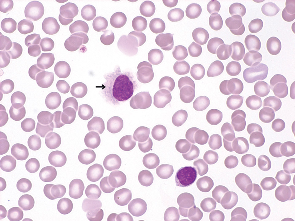
Peripheral blood (May-Grünwald-Giemsa stain) of a patient with hairy cell leukaemia. A typical hairy cell (->) can easily be distinguished from a normal lymphocyte (lower right).
<p>Peripheral blood (May-Grünwald-Giemsa stain) of a patient with hairy cell leukaemia. A typical hairy cell (->) can easily be distinguished from a normal lymphocyte (lower right).</p>
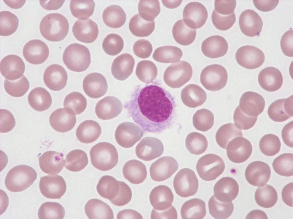
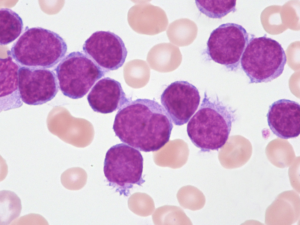
Hairy cell from a hairy cell leukaemia. Typical are the monocytic type nuclei with loose chromatin, and the grey-blue, heterogeneous cytoplasm. The hairy protrusions may be missing in some cases.
<p>Hairy cell from a hairy cell leukaemia. Typical are the monocytic type nuclei with loose chromatin, and the grey-blue, heterogeneous cytoplasm. The hairy protrusions may be missing in some cases.</p>
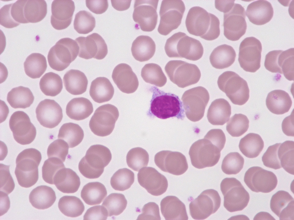
This 'hairy' lymphocyte comes from a person suffering from an active infection (without any malignant disease). The granula identify it as a T or an NK cell.
<p>This 'hairy' lymphocyte comes from a person suffering from an active infection (without any malignant disease). The granula identify it as a T or an NK cell.</p>
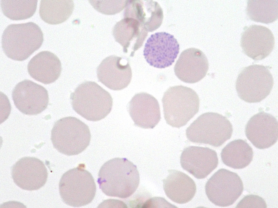
Reticulocyte staining. Characteristic HbH cell in a case of α-thalassaemia. Today, blood films are no longer investigated for HbH cells. Instead,
α-thalassaemias are diagnosed by molecular genetic tests.
<p>Reticulocyte staining. Characteristic HbH cell in a case of α-thalassaemia. Today, blood films are no longer investigated for HbH cells. Instead, </p> <p>α-thalassaemias are diagnosed by molecular genetic tests.</p>
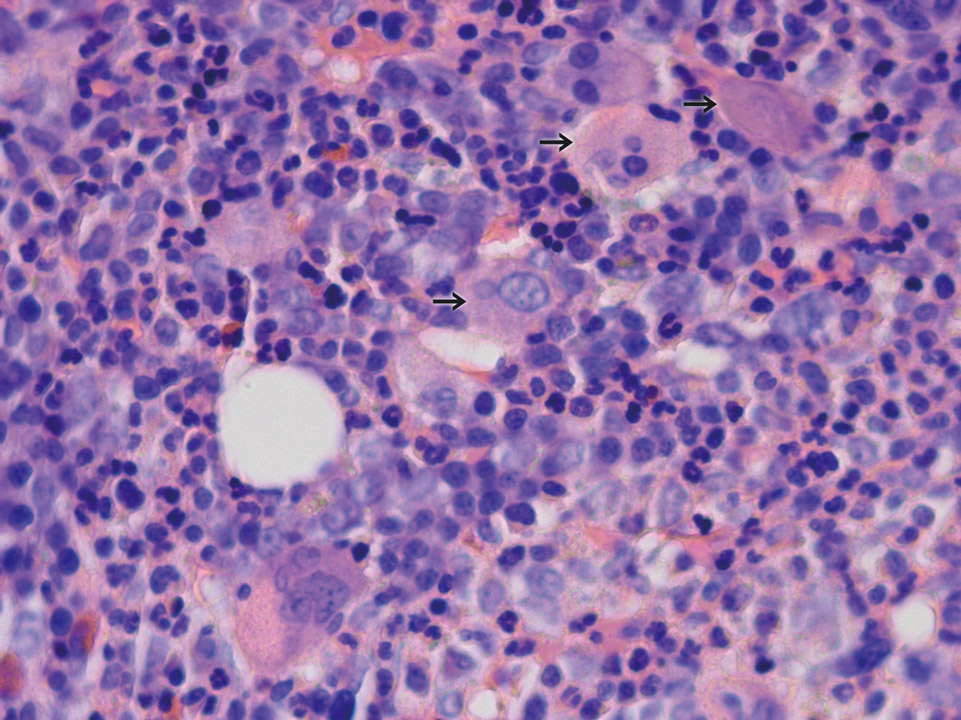
A remarkably high and conspicuous number of megakaryocytes (->) is visible in the bone marrow histology (Giemsa stain) of a patient with CML.
<p>A remarkably high and conspicuous number of megakaryocytes (->) is visible in the bone marrow histology (Giemsa stain) of a patient with CML. </p>
